 Modélisation personnalisée d'un genou par recalage d'un modèle déformable sur des données 'scanner'
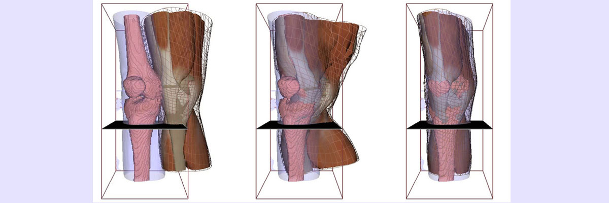
Anatomical modeling (skin and muscle)
We developed a novel approach for simulating 3D muscle deformations with complex architectures. The approach consists in choosing the best model formulation in terms of computation cost and accuracy, that mixes a volumetric tissue model based on finite element method (3D FEM), a muscle fiber model (Hill contractile 1D element) and a membrane model accounting for aponeurosis tissue (2D FEM). The separate models are mechanically binded using barycentric embeddings. Our approach allows the computation of several fiber directions in one coarse finite element, and thus, strongly decreases the required finite element resolution to predict muscle deformation during contraction. Using surface registration, fibers tracks of specific architecture can be transferred from a template to subject morphology, and then simulated. As a case study, three different architectures are simulated and compared to their equivalent one dimensional Hill wire model simulations [ART.16]. We also aim at developing a tool for predicting the behavior of soft tissues during plastic surgery procedures, we looked for the existence of homologies in the overall pattern of organization of the skin/subcutaneous tissue complex between various body parts, using high-resolution in vivo imaging methods and data available in the literature. 3T MRI scanning sequences were performed using appropriate radiofrequency coils on the face, thorax, breast, abdomen and lower extremity of six healthy volunteers. The radiological findings were segmented and converted into volumetric data. The superficial and deep adipose tissue was found to be clearly separated by an intermediate layer called stratum membranosum or superficial fascia. This continuous layer covered all the anatomical parts of the body examined. It was found to have several components in the trunk and limbs and to form a continuous layer with the superficial muscular aponeurotic system in the face. A retaining connective network consisting of superficial and deep retinacula cutis detected in all the regions investigated sometimes formed more densely packed structures playing the role of skin ligaments. The results of a 3T MRI study on subcutaneous tissue showed the existence of a common pattern of organization of the skin subcutaneous tissue complex in the various parts of the body studied. This general model is subject to quantitative variations and tissue differentiation processes promoting the sliding or contractility of the supporting tissue. Three-dimensional reconstructions were obtained by postprocessing the MRI images and will be used to perform pre-surgical simulations by settings a generic model that can be adapted to the different localization of the human body in a procedural way [ART.30, CART.39]. We also build a 3D geometric and mechanical model of the skin/subcutaneous complex (SSC) which could be adapted to the different parts of the body and to the morphological parameters of the patient. We present first the anatomical pattern of the SSC. Then, we propose a hybrid model which combines volume, membranous and uni-dimensional models. The complex internal structure of the SSC is automatically created by a procedural process. All the models are defined by some parameters which can be easily measured by medical imaging. We describe several preliminary experiments which show how this hybrid method models realistic geometrical deformations and physical behaviors and could be used for surgery simulation and planning [ART.28].