 Détection automatique des files cellulaires dans des images microscopiques de coupes de bois (collaboration avec AMAP/CIRAD)
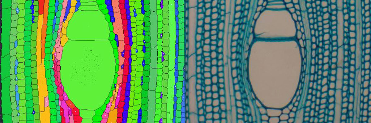
Visualization and identification : application to natural objects; Wood cell identification, tree segmentation
The analysis of anatomical sections of wood provides important information for understanding the secondary growth and development of plants. This study reports on a new method for the automatic detection and characterization of cell files in wood images obtained by light microscopy. To facilitate interpretation of the results, reliability coefficients have been determined, which characterize the files, their cells and their respective measurements. Histological sections and blocks of the gymnosperms Pinus canariensis, P. nigra and Abies alba were used, together with histological sections of the angiosperm mahogany (Swietenia spp.). Samples were scanned microscopically and mosaic images were built up. After initial processing to reduce noise and enhance contrast, cells were identified using a "watershed" algorithm and then cell files were built up by the successive aggregation of cells taken from progressively enlarged neighboring regions. Cell characteristics such as thickness and size were calculated, and a method was developed to determine the reliability of the measurements relative to manual methods. This method provides a fast, economical and reliable tool for the identification of cell files. The reliability indicator characterizing the files permits quick filtering of data for statistical analysis while also highlighting particular biological configurations present in the wood sections [ART.39]. Another of our contribution consist in an original semi-supervised method for the segmentation of in situ tree color images which combines color quantization, adaptive fragmentation of learning areas defined by the human operator and labeling propagation. A mathematical morphology post-processing is introduced to emphasize the narrow and thin structures which characterize branches. Applied in the L*a*b* color system, this method is well adapted to easily adjust the learning set so that the resultant labeling corresponds to the accuracy achieved by the human operator.