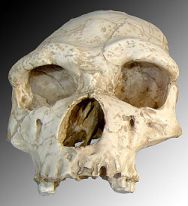
Cast of the skull of the Tautavel Man (Arago XXI)
Acknowledgment: (to be displayed when using these data)
Data provided by the ODENT Project supported by the ST2I Dept. of CNRS (http://www.lirmm.fr/ODENT/). Special Thanks to Dr. F.Dedouit, Dr. M. Faruch and all the radiological staff of the Radiology Department (head: Pr. H. Rousseau) of the Rangueil Hospital, Toulouse, France.
Warning:
All data (3D mesh or 3D image) are watermarked et therefore can be tracked.
Data can be used on a free basis but only for scientific or teaching purposes. In no case, they can be used for commercial goals.
Any use of data (e.g., scientific paper or presentation, Web site, etc.) must display:
- i. the source of the data (i.e. the address of the Web site),
- ii. the complete acknowledgement text which is given on the corresponding Web page.
Description:
Casts of the skull of the Man of Tautavel (Arago XXI) are available at the Centre Européen de Recherches Préhistoriques de Tautavel.
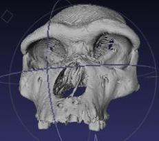
The 3D image acquisition was performed on a Siemens Sensation 16 Multi Slice Computed Tomography in the Radiology Department of the Rangueil Hospital, Toulouse, France.
- Size of the original image: 512 x 512 x 439
- Voxel size: 0.422 x 0.422 x 0.400 mm
- Image depth: 16 bits
The extraction of the bone surface mesh was performed according to the protocol described in the tutorial with the following parameters:
- MicroView:
- Image Threshold = 1000
- Surface Quality Factor = 1.00
- Enable Image Smoothing = NO
- MeshLab:
- Filters/Quadric Edge Collapse Decimation = 500000
- Filters/Clean/Remove Isolated Pieces (wrt faces num) = 400 000
- Filters/Clean/Unreferenced Vertex
- Filters/Clean/Duplicated Vertex
- Filters/Clean/Zero Area Faces
- Filters/Clean/Non Manifold Faces
The mesh in .obj format is composed of 249,846 vertices and 499,990 faces.
To download the mesh file "tautavelODENTWatermarked.zip" (5.1Mb, decompressed file "tautavelODENTWatermarked.obj": 19.5Mb), click here.
Some research work using 3D data of the skull of the Tautavel Man (Arago XXI):
- G. Guipert, M.A. de Lumley, H. de Lumley, B. Mafart. "Three-Dimensional Imagery : A New Look at the Tautavel Man" [Enter the Past]. Computer Applications and Quantitative Methods in Archaeology. Stadtarchäologie Wien (Eds). BAR International Series 1227, 2004, 100-102.
- G. Subsol, B. Mafart, A. Silvestre, M.A. de Lumley. "3D Image Processing for the Study of the Evolution of the Shape of the Human Skull: Presentation of the Tools and Preliminary Results". XIV International Congress of Prehistoric and Protohistoric Science, Liège (Belgium), September 2001. Abstract page 22 in preprints, full text in: B. Mafart and H. Delingette with the collaboration of G. Subsol (eds.) Three-Dimensional Imaging in Paleoanthropology and Prehistoric Archaeology, British Archaeological Reports International Series 1049, 37-45, 2002.
- G. Odin, G. Quatrehomme, G. Subsol, H. Delingette, B. Mafart, M.A. de Lumley. "Comparison of a Three-Dimensional and a Computerized-Assisted Method for Cranio-facial Reconstruction: Application to the Tautavel Man". XIV International Congress of Prehistoric and Protohistoric Science, Liège (Belgium), September 2001. Abstract page 23 in preprints, full text in B. Mafart and H. Delingette with the collaboration of G. Subsol (eds.) Three-Dimensional Imaging in Paleoanthropology and Prehistoric Archaeology, British Archaeological Reports International Series 1049, 67-69, 2002.
- B. Mafart, D. Méline, A. Silvestre, G. Subsol. "3D Imagery and Paleontology: Shape differences between the skull of Modern Man and that of Tautavel Man". B. Hidoine, A. Paouri (designers and directors). Video 451-452. INRIA Multimedia Scientific Communication Department, 1999.
This movie was showed at the exhibition Homo Erectus à la conquête du monde, Musée de l'Homme, Paris (France) during several months in 2000.
Contact:
- Gérard Subsol, CNRS Senior Researcher
LIRMM, Montpellier, France
[gerard.subsol@lirmm.fr]


